
Author: Mike Wong
Assessing intracortical myelin in the living human brain using myelinated cortical thickness.
AUTHORS: Rowley CD, Bazin PL, Tardif CL, Sehmbi M, Hashim E, Zaharieva N, Minuzzi L, Frey BN, Bock NA
SUMMARY: Christopher Rowley
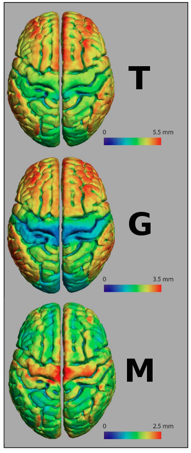
Myelin is typically associated with white matter in the brain, but many myelinated axons exist in the grey matter. The purpose of our paper was to determine if we can detect changes in this cortical myelin in disease. The distribution of myelinated axons in the cerebral cortex is regionally dependent, an observation that has been used to describe the brain’s myeloarchitecture. Since myelin is a crucial element in maintaining functional wiring and signal synchrony in the brain, it is possible that the myeloarchitecture is related to specific cor-
tical functions and may be altered if functions are altered in disease. We have developed a technique to investigate changes in myeloarchi-
tecture using MRI images with high intracortical myelin contrast. It pro-
ceeds by segmenting the brain tissue with a clustering algorithm into white matter, heavily myelinated grey matter, and lightly myelinated grey matter. By then measuring the thickness of the two grey matter tissue classes, we can detect changes in the myeloarchitecture. We have demonstrated the potential use of the technique for investigating pathologies by showing a decrease in cortical thickness in females with bipolar disorder in the middle frontal gyrus. The largest percent-
age decrease in thickness was discovered in the heavily myelinated grey matter, suggesting a decrease in the myelinated layers of the cortex. This technique allows for in vivo analysis of cortical myelin in diseased populations to uncover changes in myelin across the cerebral cortex.
CITATION: Rowley CD, Bazin PL, Tardif CL, Sehmbi M, Hashim E, Za-
harieva N, Minuzzi L, Frey BN, Bock NA (2015) Front Neurosci 9: 396.
AREA: Neuropsychiatric disease
Effects of fluoxetine and visual experience on glutamatergic and GABAergic synaptic proteins in adult rat visual cortex.
AUTHORS: Beshara S, Beston BR, Pinto JGA, Murphy KM
SUMMARY: Simon Beshara
Patching therapy is the most common treatment for children with amblyopia. For many, the acuity recovered during patching is lost when the treatment stops leaving the child with persistent amblyoia. Fluoxetine, a widely prescribed anti-depressant, has emerged as an interesting treatment option because it reinstates critical period-like plasticity and promotes recovery of vision in adult animals. Translation
of these results from animal models to the clinic, however, has been challenging because it remains unclear how this selective serotonin reuptake inhibitor affects plasticity in the visual cortex which relies heavily on glutamatergic and GABAergic synapses. An appealing hypothesis is that fluoxetine recreates plasticity by shifting synaptic mechanisms to a juvenile state. To test this we studied the effects of fluoxetine and visual experience on the visual cortex of adult rats. Surprisingly we found that fluoxetine shifted glutamatergic and GABAergic proteins to a juvenile state, but rather created a novel synaptic environment that favored mature NMDA and GABAA receptors.
CITATION: Beshara S, Beston BR, Pinto JGA, Murphy KM (2015) eNeuro (TBD).
AREA: Plasticity